
Cnidaria


The cnidarians (pronunciation "Nidarios"),(Cnidaria, from the Greek ''kníde'&# 39;, nettle) are a phylum of relatively simple diblastic animals that live exclusively in aquatic environments, mostly marine. It groups around 10,000 species, including jellyfish, polyps, corals, anemones and hydras (the only organisms that inhabit freshwater).
The name of the phylum alludes to a diagnostic characteristic of these animals, the presence of stinging cells called cnidoblasts, present in the tentacles and mouth of all members of the phylum; these function as a cavity and are in charge of keeping the cnidocytes, which are capsules where the venom is stored and are thrown by a filament towards the prey, hooking it with a sting to release the toxins. They have radial symmetry and their body plan is sac-shaped. They are the simplest animals that have nerve cells and sense organs (statocysts, ocelli).
They are an ancient group, with a long fossil history dating back, probably, to the Precambrian fauna, about 700 mya ago. However, genetic analyzes of the molecular clock of their mitochondria suggest a much earlier age for the crown group of all cnidarians, estimated at about 741 million years, long before there are any fossils that have survived to the present day.
General characteristics
Cnidarians are diblastic metazoans, that is, they are animals without tissues that have two embryonic layers (ectoderm and endoderm); although sometimes there is an analogous tissue (ectomesoderm) of ectodermal origin, not of endodermal origin like the true mesoderm of triploblastics, and from which complex internal organs never derive. Its cells are organized in two layers that act as functional units (tissues), although many cells still have some independence and some totipotency.
These organisms can be solitary or colonial individuals and sessile, sedentary or free-swimming. Some of them are suspension feeders, especially the sessile species, but most are carnivores, so they have to hunt. To carry out hunting for food, these invertebrates have specialized cells called cnidoblasts, located in their tentacles and mouth; They function as a cavity where the cnidocytes are stored, which are capsules where the venom is stored and are thrown by a filament towards the prey, hooking it with a sting to release the toxins. These cells are very sensitive, so it is enough to brush the animal's tentacles for its stingers to be expelled. In the diagram below you can see a cnidoblast and the discharge process of a cnidocyte.

Its body organization is in the form of a sack; the digestive system has a single orifice that acts as a mouth and anus at the same time, and is known as the gastrovascular cavity, where digestion takes place, it is also used as a nutrient and oxygen distribution system, and as an excretory system.
The body wall consists of:
- Ectodermis. Homologa of the ectoderm. It's the outer layer. Protective function.
- Gastrodermis. Endoderm Homologa. It is the inner layer; it delimits a wide cavity, the only of the animal, called gastrovascular cavitywhich communicates by a single orifice with the outside used for feeding, excretion and even the expulsion of genital products.
- Mesoglea. Between both layers is mesoglea, which can be very thin and acellular, or well developed and have different cell types (according to the group).
They have one or more tentacles around the mouth. The nervous system forms a network or plexus; in many groups there are non-polarized protoneurons, although there may also be polarized neurons, sensory cells, and even their grouping in sensory organs.
They tend to be polymorphic, especially colonial forms. There is no excretory system, respiratory system, or circulatory system. These functions are carried out through the gastrovascular cavity or the ectodermis.
They are essentially marine (99%). The rest are freshwater, such as the hydra, or certain jellyfish from large African lakes such as the Craspedacusta jellyfish. They are always aquatic.
They can live individually or in colonies, fixed to the substrate or free, there are even swimmers. Sometimes, part of the phases are planktonic (mobile, but carried by currents).
Approximately 10,000 species are known, varying in size from 1-2 mm to 1 m in diameter in some jellyfish, or up to 3 m in diameter in some polyps.
Color is also variable although many forms of plankton are transparent. Others are colored, and present practically all colors.
Morphology

Without paying attention to the peculiarities of each group, they appear in two fundamental forms:
Sessile form: Polyp

In general terms (see by groups for more information), it is an animal with thin walls and a wide gastrovascular cavity. Mesoglea very poorly developed. Body more or less columnar. On the aboral side (see radial symmetry) it is related to the substrate. As adults, they can be sessile or free, solitary or colonial.
They can be dioecious or hermaphrodite, and have sexual and asexual reproduction.
Variable size:
- Halamohydra: of the meiofauna, very small
- Microhydra: small and without tentacles. It is the polyp phase of the jellyfish Craspedacusta. 1-2 mm.
- Branchiocerianthus: muddy backgrounds, deep. Up to 3 m.

General diagram of a polyp (without attending groups)

General diagram of a jellyfish (without attending groups)


Free Form: Medusa

1.- Ectodermis; 2.- Mesoglea; 3.- Gastrodermis; 4.- Stomago; 5.- Radio channel; 6.- Circular channel; 7.- Tentacle; 8.- See it. 9.- External nerve ring; 10.- Internal nerve ring; 11.- Gónada; 12.- Manubrio; 13.- Boca; 14.- Exumbrela; 15.- Upload it..
It has two perfectly defined surfaces; one concave where the mouth is located (oral side, see radial symmetry) and another the opposite (aboral). The set is known as umbrella, where the exumbrella (aboral) and subumbrella (oral) are differentiated.
The mouth is prolonged into a manubrium. Tentacles can come out of the handle (or not, depending on the group). In addition, the umbrella may elongate into umbrellar tentacles (which may or may not have a tentacular cavity, an extension of the gastrovascular cavity).
The mouth opens into the gastrovascular cavity from which radial canals branch out towards the walls of the jellyfish. The umbrellar canal encircles the entire jellyfish. If the tentacles are hollow, the tentacle canal also appears.
The entire cavity (including the channels) is lined by gastrodermis. The rest by ectodermis. There is highly developed mesoglea in the exumbrella, while in the subumbrella it is very poorly developed.
Variable size, from small jellyfish with colonial polyps such as Obelia (1-2 mm) to large jellyfish such as the nomura that can reach 2 m in diameter and weigh up to 220 kilograms. Giant jellyfish are mythical animals.
Internal anatomy
Let's look at the cell types that appear in cnidarians:
Ectodermis

The ectodermis consists of various types of cells:
- Cells mioepiteliales or epitheliomusculares. The fundamental type is a cylindrical (meduse and polyp) cell with two defined parts, an apical epithelial and another contrable basal that has linens of actin and myosin. They have double function, coating and contraction.
- Interstitial cells or Baseline. They are at the base of myoepithelial cells, arranged as a wedge, and have a very large nucleus. They are very rich in ribosomes, and are totyped cells, so they can originate any other cell type.
- Cnidocitos or cnidoblasts. The cnidocytes are scattered throughout the epidermis, housed or invaded in myoepithelial cells, especially in the tentacles. It is a very specialized type of cell, exclusive of the cnidaries. They have a stiff cyllium.cnidocilio) which is a stimulus receptor; in the basal part it has extensions that contact other cells or the nervous system (cnidopodios); inside this cell there is a large vacuola (cnido or nematocyst), within which is the hilarious filament (the shooting of this filament is the "picture" of the jellyfish, polyps, anemones, etc.).
- Neurons. Nervous cells, unpolluted (protoneurones) and other polarized cells, which can be bipolar or multipolar. The nervous system forms a plexo that is placed under the epidermis.
- Sensory cells. Elongated cells with the rounded apex end or provided with a sensitive slime and the basal zone provided with one or more extensions that connect with the neurons
It is not clear whether the myoepithelial cells rest on a true basal lamina, like the true epithelial tissues of other metazoans, although the mesoglea itself can be interpreted as the basal lamina of cnidarian epithelium.
Mesoglea
Gelatinous substance that separates the two epithelial layers. It can vary from a thin, noncellular membrane to a thick gelatinous layer with or without wandering amebocytes. Presents abundant collagen and pre-collagen. Contains numerous cells, such as scleroblasts.
Gastrodermis
- Muscular nutritious cells or mioepiteliales gastrales. They remind the myoepithelial cells of the epidermis, but they are higher and narrower, with the less developed contráctil part in the base, without prolongations and with 2-5 scourges on the apical side that it uses to move the fluids of the gastrovascular cavity. This cell is characterized by the dual possibility of pinocytosis and phagocytosis. Collect partially digested materials from the gastrovascular cavity and pass them to the rest of cells. Another characteristic is that its connective part is perpendicular to that of the ectodermis.
- Secret cells. They are secretive cells of apocrine type, high, some flagellates, in which many substances are produced (mucopolisaccharides, mucoproteins, digestive enzymes, etc.). Segregan substances that lub and digest foods that enter the gastrovascular cavity.
Among them, there are sensory cells (in fewer numbers than in the ectodermis) and basal cells. cnidocytes may also appear. Poorly developed nerve plexus.
Nerve endings
In some groups there are receptors of various kinds (tactile, chemical, light, gravitational, etc.)
Touch receptors
Tactile receptors are hair cells with typical processes like those already seen in internal anatomy.
Photoreceptors

Receptors for light stimuli
The simplest are the so-called eyespots, or eyespots, which are pigmented spots that appear in certain jellyfish in the umbrella. Made up of groups of cells of two types: ciliated sensory cells and cells inside which a pigment (rhodoxin) accumulates. The pigment is located at the base of the cilium symmetrically or asymmetrically, in a folded membrane. The pigmented cell sends information to the typical sensory.
More advanced are the ocelli, which are a small recess in the epidermis in which the photoreceptor cells are in the center, surrounded by pigmented cells.
This ocellus becomes complicated into a cup-shaped ocellus, in which there is a large recess, at the bottom of which are the photoreceptors and surrounding the pigmented ones.
A little further on, in some jellyfish, the distal part of the cell is detached and vacuolar extensions are formed that fill the slit, the "cup", forming a kind of lens.
These receptors do not allow them to distinguish objects. only lights and shadows. This is what we call encephalitis.
Organs of balance

Various organs of balance have also evolved.
States are structures that have rows of two types of cells, some ciliated sensory cells, and others (lithocytes) that accumulate inside a calcareous ball (statolith). If the animal rotates, as the cell with the statolith hangs by gravity and weighs, it moves and touches one of the surrounding sensory cells. Thus the animal is kept informed of its position.
Derived from this, we have statocysts, with a larger cleft that may even be closed and not communicate with the umbrella. In other cases, the statoliths are detached from the lithocytes or foreign particles are used for the same function.
There are also statorabdos, which are small tentacles with one or more statoliths. This tentacle hangs down surrounded by sensory cells.
Clothes
In the order Scyphozoa there is a highly developed organ, the ropalia, which is a chemostate-photoreceptor center. It is the most complex sensory structure of cnidarians, and it only appears in the motile phase. The most complex ropalia of all is found in the order Cubozoans.
Toxins in a bite
Mechanical and chemical stimulation of the beards surrounding the cnid causes a calcium-mediated bioelectric signal to allow the nematocyst to come to the surface to express the venom. The exit of the nematocyst occurs in 3 milliseconds and penetrates to a depth of 0.9 mm, depositing the toxin in the microvasculature of the dermis, to then be absorbed into the systemic circulation while it is anchored to the victim.
The toxic fraction of the fluid contained in cnidocytes is a complex mixture of substances of a chemical nature, such as catecholamines, bioactive amines (histamine, serotonins), bradykinins, collagenases, hyaluronidases, proteases, phospholipases, fibrinolysins, dermatoneurotoxins, cardiotoxins, myotoxins, nephrotoxins, neurotoxins and protein antigens. The protein component of the toxin tends to be heat labile, not dialyzable, and is degraded by proteolytic agents. In addition to proteins, various enzymes, amines, carbohydrates, and lipids have also been found. All of these substances can have neurotoxic, cytotoxic, sometimes hemolytic, and cardiotoxic effects. The toxins vary depending on the different types of cnidocytes and within these they can also differ according to the species, since there are very light poisons and also others such as that of the sea wasp, which can cause death.
The toxin causes alterations in the transport of sodium and calcium channels, breaks down cell membranes, releases inflammatory mediators, and acts as a direct toxin in the myocardium, nervous tissue, liver, and kidneys. It can also produce strong itching in the person's kidney area.
Once the bite has occurred, care must be taken not to scratch or rub the area where the burning sensation or intense pain is noticeable. Do not wash with fresh water, since the osmotic change causes more cnidocysts to shoot, but with salt water. Cold compresses should be applied as soon as possible for 5-15 min. These compresses can be prepared with a plastic bag filled with ice, and the piece of ice should not be applied directly to the skin, as it would have the same effect as fresh water. The application of hot compresses is contraindicated, since the heat favors the systemic absorption of the venom.
Function of some toxins
Venoms are going to be divided into 4 groups, which are enzymes, neurotoxins, cytolysins and non-protein bioactive components. The enzymes will work like a virus, attacking the cells, infecting them in order to produce many more enzymes that attack the rest. Cytolysins will work very similar to enzymes, the only difference is that they will attack more specific parts of the victim's body, such as some of the systems. Neurotoxins are capable of attacking the central and peripheral nervous system mainly, this damage will cause a series of damage to the systems, in addition, many can be degenerative in the part where the bite was carried out. Finally, the bioactive components will be compounds that are a little more painful in the area of their bite or that enhance the venom, despite this, they become a little less aggressive towards the rest of the toxic substances.
The venom of these cnidarians will contain different types of toxins with different effects, such as
- Palitoxine: is an intense vasoconstrictor and is considered one of the most poisonous non-protein substances. In the image next door we observe the structure of this toxin.
 Palitoxin: the second most potent non-protein toxin known on Earth. It was discovered in 1971 in cnidarios
 - Hypnocin: causes drowsiness, muscle and respiratory paralysis and has anesthetic properties.
- Thalassina: produces urticaria and edema in small doses. At high doses it can cause nervous paralysis, cardiac arrest and even death.
- Congestina: causes digestive tract congestion, vomiting, abdominal pains, decreased blood pressure and respiratory problems.
- Tetramina: possibly with paralysing action.
- 5 hydroxytriptamine: responsible for the pain and release of histamine.
- Actinoporins: Multigenic family of toxic proteins produced by various species of anemones. These proteins act by making pores in the plasma membranes of the cells. In Scheme 3 you can see a representation of a pore formed by actinoporins Actinia fragacea sp.
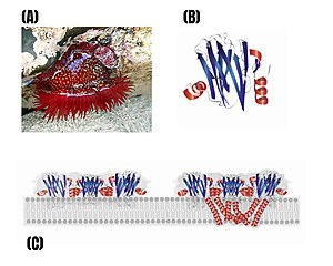 Pore model consisting of actinoporins. (A): photo Actinia fragacea sp. (B): Crystalline structure of the C-toxin frigacea in a monomeric state. (C): model of the transmembrane pore of fagaceatoxine C.


Cycle

The cycle varies a lot between groups, so for more information see the different classes. Although it is characterized by the alternation of generations.
In general, the egg has little yolk and undergoes total and equal segmentation. In many, gastrulation is by delamination to reach a general larval state, the planula larva, ciliated, swimming, which will look for a place to fix itself in the substrate, giving rise to the polyp, which grows and at a certain time, by agametic reproduction (usually budding) by a process called strobilation, gives rise to jellyfish, in which the gametes that will form the new egg will mature.
This complete cycle is a metagenetic cycle. Within the groups there are deviations; For example, there are polyps that give rise to polyps (Hydra by budding creates a polyp that grows and separates). There are also jellyfish whose egg develops into a planula, but which will develop into a jellyfish very quickly. There are also intermediate phases; siphonophores are colonial, and there are polypoids (derived from polyp) and medusoids (derived from medusa) coexisting in the colony. All these cycles that are not complete are called hypogenetic.
Classification
The traditional classification of cnidarians recognizes four classes and assumes that the anthozoans are the most primitive class and from which the others have been derived:
Anthozoa (Anthozoa)
Only polyps (anthopolyps), known as anemones and corals. There is no jellyfish form. The polyp gives, by asexual or sexual reproduction, polyps. There are cnidocytes in the gastrovascular cavity, sometimes very powerful, even in filaments protruding through the body wall. The endodermal gonads. The gastrovascular cavity is completely divided (the septa come from the gastrodermis and the mesoglea). The oral side enters the gastrovascular cavity and gives rise to a stomodeum or pharynx.
Cubozoa (Cubozoa)

They only have jellyfish (cubomedusas), cubic in shape, with special, very powerful cnidocytes. Until recently it was considered to be an order of the Scyphozoa. They are characterized by their tetrameric division, which separates the gastrovascular cavity into four bags. The umbrella edge is not scalloped and the margin of the subumbrella folds inward to form a velarium.
Hydrozoans (Hydrozoa)
They have hydropolyps and hydromedusae. Jellyfish are veiled (they are craspedo jellyfish). They do not have cnidocytes in the gastrovascular cavity. The gonads are always of ectodermal origin. In a cross section, the gastrovascular cavity is single, undivided. The mesoglea of the polyps is poorly developed.
Scyphozoans (Scyphozoa)
It has a small and inconspicuous polyp (scyphopolyp or scyphostoma) and medusa (scyphomedusa ), which lacks a veil (acraspedota). They present cnidocytes in the gastrovascular cavity. The gonads are endodermal. Once matured, however, they can be stored in the endodermis. Its gastrovascular cavity is incompletely divided by 4 incomplete partitions or septa in interradial position (see radial symmetry); the septa separate 4 gastral pouches.
Currently, the World Register of Marine Species also includes the following classes in Cnidaria:
Mixozoans (Myxozoa)
They are a class of microscopic parasitic animals, long classified as protozoa within the sporozoans. Parasitism occurs by spores, with valves that would contain one or two sporoblasts and one or two capsules with filaments that would anchor the spore to the host. In 2015 a study revealed that myxozoans are actually extremely small cnidarians with a simplified genome.
Polypodiozoa (Polypodiozoa)
It is a monospecific class of parasitic cnidarians. It is one of the few cnidarians that live inside the cells of other animals. Molecular data suggest that it could be related to myxozoans (also parasites), but this point is controversial, so its classification is temporary, since its phylogenetic relationships are unclear.
Staurozoans (Staurozoa)
These are sessile forms. The aboral surface, corresponding to the exumbrella of other jellyfish, is prolonged into a stem thanks to which they are fixed to the substrate, especially algae and rocks. Until recently they were considered an order within the class Scyphozoa, but were raised to class status after cladistic study.
Phylogeny
Many phylogenetic theories have been proposed to relate the different groups of cnidarians. Most of the authors defend that the possible pre-cnidarian was a planuloid organism adapted to sessile life, acquiring radial symmetry, which would have undergone an invagination that later gave way to the gastrovascular cavity. Was the pre-cnidarian shaped like a medusa or a polyp? There are 2 theories:[citation needed]
| Polypoid theory Defends that pre-cnidary was a polyp
| 1 Radial symmetry, cnidocytes, gastrovascular cavity with partitions, endodermal gonades, tentacles around the mouth, polypoid-shaped body, circular mitochondrial DNA; 2 cnidocytes with cnidocil, linear mitochondrial DNA; 3 medusa acraspedota; 4 Clothes; 5 metagenetic cycle with reduction of the polyp phase; 6 estrobilization; 7 cubic umbrela; 8 guard; 9 medusa craspedota; 10 loss of gastrovascular cavity tabication; 11 weight loss of mesogle and reduction of the amount of amoebocytes; 12 ectodermals. | |||||||||||||||||||||||||||
| Medusoid Theory Defends that pre-cnidary was a jellyfish
| 1 Radial symmetry, gastrovascular cavity without tabicar, ectodermals, tentacles of the mouth, larva plánula that becomes the actinula larva; 2 polyp, metagenetic cycle; 3 endodermals; 4 gastrovascular cavity with partitions; 5 reduction or loss of the polyp phase; 6 Clothes; 7 estrobilization; 8 Cubic umbrela, guardio; 9 loss of the medusa phase; 10 hexarradial or octorradial symmetry; 11 faringe; 12 sifonoglifos; 13 cnidocytes without cnidocil or opérculo. |
Studies based on molecular phylogeny corroborate the monophyly of cnidarians and that the polyp form probably preceded the medusa form in cnidarian evolution. It also suggests that the cnidarians are made up of two groups that could have the category of subphyla, the anthozoa and the medusozoans; the latter group the cubozoans, the scyphozoans, the hydrozoans and the stauromedusas, so the most recent genomic relationships give the following result:
| Cnidaria |
| ||||||||||||||||||||||||||||||||||||||||||
Contenido relacionado
Illicium
Category:Molecular biologists
Philadelphia chromosome