Central Nervous System
The central nervous system is one of the portions into which the nervous system is divided. In vertebrate animals, it is made up of the brain and the spinal cord, it is covered by three membranes: dura mater (external membrane), arachnoid mater (intermediate), pia mater (internal membrane), generically called meninges and protected by bone envelopes, which are the skull and vertebral column respectively.
This is a very complex system, since it is responsible for perceiving stimuli from the outside world, processing information, and transmitting impulses to nerves and muscles. The nervous system of vertebrate animals, including mammals and man, can be divided into two distinct parts, the central nervous system, made up of the brain and spinal cord, and the peripheral nervous system, made up of sensory and motor nerves. that link the central nervous system with the rest of the organism.
Structure
The central nervous system consists of the brain and spinal cord.
- The Right. is the part of the central nervous system that is protected by the skull bones. It is formed by the brain, cerebellum and encephalic stem.
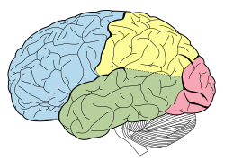
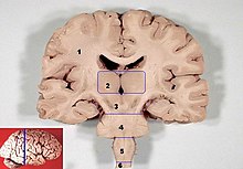
- The brain is the most voluminous part. It is divided into two hemispheres, one right and one left, separated by the interhemispheric scent and communicated by the silent body. The surface is called cerebral cortex and is formed by folds called circumvolutions, constituted of gray substance. Subyacent to it is found the white substance. In deep areas there are areas of gray substance forming nuclei such as thalamus, nucleus and hypothalamus. Each brain hemisphere has several cysuras that divide the cerebral cortex into lobes:
- Front lobe. It is located in previous position.
- Temporary lobe. It is located in a lateral position behind the frontal lobe.
- Parietal lobe. It extends on the outer face of the hemisphere, under the temporal lobe.
- Occipital lobe. It is located at the back of the brain.
- The brain is the most voluminous part. It is divided into two hemispheres, one right and one left, separated by the interhemispheric scent and communicated by the silent body. The surface is called cerebral cortex and is formed by folds called circumvolutions, constituted of gray substance. Subyacent to it is found the white substance. In deep areas there are areas of gray substance forming nuclei such as thalamus, nucleus and hypothalamus. Each brain hemisphere has several cysuras that divide the cerebral cortex into lobes:
- The ceebelo is at the bottom and back of the brain, housed in the posterior brain pit next to the brain stem.
- The Encephalic stem composed of mesencephalo, anular protuberance and the rachid bulb. Connect the brain with the spinal cord.
- La spinal cord It is an extension of the brain, as if it were a cord that extends through the inside of the spine. In it the gray substance is found inside and white on the outside. In the spinal cord the reflex arches are established.
| Central nervous system | Right. | Cerebro |
| Cerebelo | ||
| Encephalic stalk | ||
| Spinal cord | ||
Brodmann Areas
In 1878, Korbinian Brodmann carried out a study of the cerebral cortex and divided it into 52 different areas according to their location. It has been proven that many of these areas have a specific function, for example area 17 located in the occipital lobe corresponds to the primary visual cortex and is where nerve impulses from the optic nerve are processed, areas 44 and 45 are called areas of Broca, and are related to language.
Frontal lobe
| Side vision of the cerebral lobes. |
It is located in the anterior part of the brain, its size corresponds to approximately one third of the cerebral cortex. Evolutionarily it is one of the most modern parts of the brain and is highly developed in the human species. The Rolando fissure separates the frontal lobe from the parietal lobe behind it, while the Sylvian fissure serves as a boundary with the underlying temporal lobe.
Its functions are of great importance, within the frontal lobe is the primary motor area that is in charge of issuing orders to carry out movements of all voluntary muscles and Broca's area related to the production of language. Its neural circuits are closely related to the ability to reason, solve complex problems and abstract thinking.
Parietal lobe
The parietal lobe is part of the cerebral cortex, it is located behind the frontal lobe, separated from it by the Rolando fissure. In its posterior portion it comes into contact with the occipital lobe, while the Sylvian fissure separates it from the temporal lobe located below.
In the parietal lobe is the somatosensory area that captures and processes sensations of touch, pain, and temperature throughout the body. When there are lesions that affect the parietal lobe, a symptom called asomatoagnosia may occur, which consists of that the patient is not able to recognize parts of his body, such as a lower or upper extremity, which can be a cause of great concern and concern.
Temporal lobe
The primary auditory area that receives and processes information from the ear is located in this lobe. Therefore, a lesion in the temporal lobe can cause partial deafness even though the ear and auditory nerve are not damaged. Next to the previous one is the secondary auditory and association area, which includes Wernicke's area, which is very important in linguistic function and word comprehension.
Occipital lobe
The occipital lobe is smaller than the anterior lobe and is located in the posterior region of the brain, separated from the cerebellum by the dura mater. Contains the primary visual cortex that receives information from the retina via the optic nerve. Neurons in the primary visual cortex are responsible for processing visual stimuli and interpreting shapes, movement, and other aspects of vision. Therefore, when there are lesions that affect the occipital lobe, cortical blindness can occur, which is characterized by the fact that the person cannot see although the eye does not show any apparent damage.
Corpo callosum
The corpus callosum is an important structure of the brain that is made up of fibers that act as a communication channel between the right and left cerebral hemispheres, so that both work together and complement each other.
Inner capsule
The internal capsule is a thick set of both ascending and descending nerve fibers that communicate the cortex with the lower regions of the central nervous system, the fibers are of diverse origin, but many of them carry motor or sensory information. On their way they pass near the region of the thalamus and the basal ganglia. The internal capsule is a very sensitive region, any injury to this area damages numerous nerve fibers and consequently causes severe neurological deficits.
Thalamus
The thalamus is a portion of the brain located above the brainstem, almost in the center of the brain. It is about 3 cm long and is made up of gray matter, that is, the soma of neuronal cells. It fulfills the function of a relay station for nerve signals and an integration center where sensory impulses are processed before continuing their journey to the cerebral cortex. It also receives signals that follow the opposite direction and reach the thalamus from the cerebral cortex.
Hypothalamus
The hypothalamus is a small region of the brain made up of gray matter. It is located immediately below the thalamus. It is about the size of an almond and performs important functions, including linking the nervous system with the endocrine system through the pituitary gland.
Basal ganglia
The basal ganglia should actually be called the basal ganglia as they are not true ganglia. They are brain structures formed by neuronal bodies (grey matter) located at the base of the brain. They are made up of different nuclei: caudate nucleus, putamen, globus pallidus, nucleus accumbens, lenticular nucleus, striatum, cerebral amygdala, and substantia nigra. For many years it has been considered that the function of the basal ganglia is solely the control of body motility, however, it has been proven that they play an important role in other functions such as learning and memory. The functional alteration of the basal ganglia causes Parkinson's disease.
Grey matter and white matter
The cells that make up the central nervous system are arranged in such a way that they give rise to two very characteristic formations:
- Gray substance, formed by the soma of the neurons and their dendrites, also by amylinical fibers.
- White substance, formed mainly by myelinized nerve prolongations (axons), whose function is to drive the information, through nervous impulses to other neurons. The color of the white substance is due to the myelin of the axons.
Cerebrospinal fluid
The central nervous system has cavities called cerebral ventricles in the brain and an ependymal canal in the spinal cord. These spaces are filled with a clear, colorless fluid called cerebrospinal fluid. Its functions are very varied: it serves as a means of exchange for certain substances, as a system for eliminating residual products, to maintain the proper ionic balance, and as a mechanical damping system.
The cerebral ventricle system is made up of two lateral ventricles that are located symmetrically and are connected to the third ventricle, which through Silvio's aqueduct communicates with the fourth ventricle.
Embryonic development
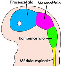
The central nervous system of vertebrates develops as a hollow tube produced by the union of 2 ectodermal folds. The spinal cord retains this tube structure, while in the brain the tube widens into 3 primitive vesicles that They are called the prosencephalon (forebrain), the midbrain (midbrain), and the rhombencephalon (hindbrain). Subsequently, these 3 vesicles become 5 by dividing the forebrain into diencephalon and telencephalon and the rhombencephalon into metencephalon and myelencephalon. These 5 primitive vesicles give rise to all portions of the adult brain, according to the following scheme.
| Prosencéfalo | Telencéfalo | Surface, tonsil, hippocampus, neocortex, side ventricles | |
| Direct it. | Epithalamus, thalamus, hypothalamus, subthalamus, pituitary gland, pineal gland, third ventricle | ||
| Mesencéfalo | Téctum, cerebral peduncle, pretectum, Silvio aqueduct | ||
| Break it up. | Metencéfalo | Logencephalic bridge, cerebellum | |
| I'm sorry. | Bulbo raquíd | ||
The central nervous system is covered in the embryo by a single meninge: the primitive meninge. This is the only membrane that develops in fish. It then divides into the outermost dura mater and a secondary, innermost meninges, the piarachnoid mater, and eventually divides into the pia mater and arachnoid mater.
Diseases
Infections
The central nervous system can be the target of infections, coming from four main routes of entry: dissemination through the blood, which is the most frequent route, direct implantation of the germ due to trauma or iatrogenic causes, local extension secondary to a local infection and the peripheral nervous system itself, as occurs in rabies.
Encephalitis and myelitis
Encephalitis is an acute diffuse inflammatory process that causes neuronal death, generally of infectious origin. Although there are many possible causes, one of the most common is the herpes virus (herpetic encephalitis).
Meningitis
Meningitis is an inflammation or infection of the meninges, either leptomeningitis that is centered in the subarachnoid space, or pachymeningitis that is centered in the dura mater. Pyogenic meningitis is caused by bacteria, mainly: Haemophilus influenzae, Neisseria meningitidis and pneumococcus.
Neurodegenerative diseases
- Multiple sclerosis: disorder characterized by episodes of recurrent neurological deficit with demylinization by auto-inmunitory or inmunitary mechanisms. It appears at any age, although it is rare in childhood or after 50 years of age, affecting women at a 2:1 ratio for men.
- Alzheimer's disease: is the most common of neurodegenerative diseases and the first cause of dementia, of sporadic appearance, although 5-10 % are of a family character and the incidence increases with age, becoming greater in people over 85 years of age. It is characterized by a lack of progressive memory due to degeneration of the cortex, of temporary and parietal association, causing also affective disorders.
- Parkinson's disease: they affect the basal ganglia by producing a movement disorder, appreciating stiffness and slowness in the voluntary movements (bradynamics) and tremor of rest.
- Huntington's disease: a disorder of choreiform-type movements and dementia in patients between 20-50 years with a genetic factor of autosomal inheritance dominant by a causative gene located in the short arm of chromosome 4.
Central Nervous System Tumors
In general, the frequency of intracranial tumors is between 10 and 17 per 100,000 inhabitants. Approximately half are primary tumors and the rest are metastatic. Central nervous system tumors derive from various tissues, so they are divided into neuroepithelial and non-neuroepithelial.
Neurepithelial tumors are a group of primary brain tumors called gliomas. They derive from astrocytes, oligodendrocytes, ependyma, choroid plexuses, neurons, and embryonic cells. They usually diffusely infiltrate the adjacent brain, making surgical resection difficult. The most frequent are: astrocytoma, oligodendroglioma, ependymoma, neuroblastoma and medulloblastoma.
Non-neuroepithelial tumors can be of several types: meningioma, schwannoma, also called neurinomas, primary brain lymphoma, and germ cell tumor.
Contenido relacionado
Adrenocorticotropic hormone
Endocardium
DOPA
Human physiology
Huntington's disease


