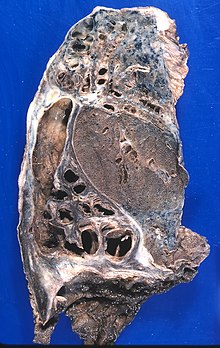
Bronchiectasis
Bronchiectasis is an abnormal and irreversible dilation of the bronchial tree, responsible for conducting air from the trachea to the functional respiratory unit (pulmonary alveoli), which can be localized or generalized. It was first described in 1819 by the French physician René Laënnec.
Etiology
Bronchiectasis is understood to be a congenital or acquired chronic disease. In the particular case of congenital bronchiectasis, this derives from the agenesis (a concept that in simple terms, is applied in the case of incomplete or imperfect development of an organ or its parts) of the alveoli with cystic dilatation resulting from the bronchi. terminals.
The etiology of acquired bronchiectasis is difficult to decipher. The development of this can occur at any age, with a higher incidence in early childhood. In general cases, it develops after a bronchial infection, in most cases severe childhood pneumonia, (commonly as complications of measles, immunological deficiencies, foreign body aspiration or bronchial erosion) that destroys the elastic and muscular supporting components. of the bronchial wall. It is a common finding in the pulmonary manifestations of cystic fibrosis.
Note that the incidence of bronchiectasis has dramatically decreased with the widespread use of antibiotics and pediatric immunizations. In adults, where the disease occurs in lesser numbers, bronchiectasis may develop after destructive inflammatory bronchial changes, necrotizing pneumonia, or lung abscess.
The development of the bronchi during the course of the disease is characterized in that they are generally reduced in length, widened, sometimes chronically inflamed and, on occasions, they are described as bronchiactatic, which leads us to believe that it is a characteristic typical of the pathology.
Pathology
Bronchiectasis can be unilateral or bilateral, and is more common in the lower lobes. It is characterized by presenting itself in three different forms: cylindrical, varicose or saccular.
Cylindrical bronchiectasis presents slightly dilated bronchi and bronchograms show them with a beaded appearance where they end abruptly without the usual branching shape. The smaller distal bronchi can be identified pathologically but are filled with inflammatory exudate.
In varicose bronchiectasis, the bronchi are irregularly dilated and contracted.
And finally, saccular bronchiectasis, also called cystic bronchiectasis, presents that the dilated bronchi are much wider and tend to be saccular (sac-shaped), with only the first ramifications of the tracheobronchial tree being present.
The bronchial walls microscopically show extensive signs of inflammatory destruction, chronic inflammation, secretion, and loss of cilia, and there is extensive destruction and disappearance of adjacent interstitial and alveolar areas. These areas of parenchymal destruction can give rise to extensive areas of increased organization and fibrosis, as well as decreased volume.
It can be congenital, although in most cases it is acquired during life for various reasons:
- Pulmonary tuberculosis.
- Respiratory infections in childhood: Infection is the most common cause. Adenovirus, which originates the common cold and other conditions, and the influenza virus, are the main agents that produce it. Also bacterial infections. Other frequent causes are bronchial obstruction (by aspiration of foreign bodies in children or benign tumors in adults).
- Cystic fibrosis.
- Immunodeficiencies.
- COPD, asthma.
Sometimes they are congenital and accompany other malformations, pulmonary or not.
- Multiple other less frequent causes.
It generally begins in childhood, although it can have a silent evolution for years or with trivial symptoms of bronchitis. They are more common in young people. In the elderly they are secondary to other diseases. They are more common in males.
The different processes that occur in the organism when this disorder occurs are the destruction of the structural components of the bronchial wall, accumulation of thick secretions, sometimes purulent, that close the most peripheral airways and alterations in the bronchial vascularization (production or increase in the number of vessels) that can cause hemoptysis of different severity.
You can find risk factors that contribute to its triggering such as sinusitis and mouth infections, from which bacteria can be aspirated; by aspiration of gastric content, which carries germs and irritates due to its acidity, or toxic gases capable of producing inflammation. Also due to the infection of the fungus called Aspergillus, which produces an exaggerated immune response.
In clinical practice, a classic triad of symptoms is observed.
- Chronic expectations.
- Recurrent episodes of respiratory infection.
- Recurrent episodes of hemoptisis.
Treatment for bronchiectasis:
- Apply antibiotics and physiotherapy. Physical therapy plays a very important role, but it should not be applied when there is hemoptoic sputum.
Contenido relacionado
Glaucoma
The New England Journal of Medicine
Come to

