Ascomycota
The ascomycetes (Ascomycota) are a division of fungi with septate mycelium that produce endogenous ascospores. They can be unicellular and thallophytes. They have been isolated from extreme places, from inside rocks on the Antarctic ice plain to the depths of the sea. They exist in terrestrial and aquatic environments, on substrates such as wood, keratin materials (nails, feathers, horns, and hair), dung, soil, and food, among others. It includes a great diversity of types of fungi, such as "false mushrooms", truffles, "terrestrial tongues" among others that form ascocarps, most lichens, some molds of bread and fruits such as Penicillium from which penicillin was obtained, the classic yeasts (Saccharomycetes) used in the fermentation of beer and other foods. Like other fungi, it also includes animal and plant parasites, including those that cause dermatophytosis or ringworm in humans, such as athlete's foot.
It is the largest taxonomic group with 32,000 described species in 3,400 genera. It is worth mentioning that this type of fungus can reproduce asexually by means of haploid spores called conidia; however, the name of the group comes from the Greek (askos) which means bag since these organisms are characterized by developing in their sexual phase structures called ascas that are similar to a bag.
Playback
Asexual reproduction
Asexual reproduction is carried out by conidia-type aplanospores, chlamydospores. They are the basics; members can be found that possess types of those spores. The basic difference between the types of spores is:
Conidia: Certain hyphae by non-symmetric mitotic division (by non-symmetric mitosis, large daughter cells and small daughter cells). The strangulation is completed and a first spore is formed, the content of this spores and another spore is formed by the same procedure, as a result we have hyphae with a large number of spores. The distal cell maintains its shape, then the cells that act as spores. The resulting protoplasm stretches to the original size of the portion and again forms another spore cell.
Oidium : Any vegetative hypha (it is fragmented in each septum) can originate; without previous transformation of the hyphae, the septa are formed, the hyphae are separated and each piece of segment acts as a spore, being able to preserve in some cases the morphology it had and in others round off this characteristic that most pathogens possess.
Chlamydospora: In vegetative hyphae, mainly in the distal portion, the protoplasm of each cell contracts, is covered with a thick wall, and when the maternal walls disintegrate, they come out.
Sexual reproduction
Morphogenesis of Asci (Ascogenesis)
In general terms, the beginning of its sexual phase begins with the recognition and sexual differentiation of its hyphae. Once this is done, the specialized hyphae carry out plasmogamy, which consists of fusing their cytoplasms, allowing the two haploid nuclei to be compartmentalized in the same cytoplasm (dikaryon). Subsequently, karyogamy is performed, which consists of the fusion of these two haploid nuclei, becoming a single diploid nucleus. The last step is meiosis, where the crossing over of genetic material occurs, resulting in haploid nuclei that, through sporogenesis, form haploid spores of sexual origin called ascospores.
Within the Ascomycota division some homothallic organisms can be found, in which any male sexual organ is compatible with any female sexual organ of the same species; While most of the group are heterothallic, that is, they have two reproductive types, which are designated as (A1) and (A2), for the sexual organs to carry out copulation, two opposite mycelia must be present., where the ascogonia and antheridia do not come from the same mycelium.
Ascomycota plasmogamy is carried out through different mechanisms such as: gametangial contact where the gametangia come into contact from a tricogyne and present a bridge to allow the flow of nuclei; gametangial copulation, which is characterized by giving way to plasmogamy from the fusion of gametes; spermatization: which is the union of a sperm to a receptor structure or somatogamy which is characterized by the fusion of somatic hyphae; however, gametangial contact predominates in this group.
In yeast and some fungi, asci originate directly from a cell; however, in most ascomycetes it develops from specialized hyphae such as the ascogenous hyphae, which is frequently multinucleated. The development of the asci begins with the division of both types of nuclei (male and female) contained in the ascogenous hypha and simultaneously a fold is formed that goes from the tip of the hypha to the stem, generating a structure similar to a "hook". ”. Subsequently, two septa are formed that separate the nuclei and distribute them, leaving the terminal cell uninucleate and the penultimate cell binucleate (male and female nucleus) and as the tip of the hook fuses with the stem, and the penultimate cell n+n (dicaryont) begins to elongate and at the same time, their nuclei fuse (karyogamy) and it becomes diploid (2n). It divides by meiosis and generates four haploid meiospores (originating from meiosis) and divides again by mitosis until finally eight ascospores are obtained.
It should be noted that the asci in formation have a continuous double membrane system with the endoplasmic reticulum (ER), so that each nucleus and a portion of cytoplasm will be surrounded by a double membrane, once this occurs the formation of the asci primary wall of the ascospore and then the other layers of the wall. On the other hand, it has been seen that the septa that are responsible for distributing the genetic load in the asca are different from the Woronin bodies and are highly specialized structures that they block the pores of the septum at the base of the ascus. It is likely that this specialization of the septa allows a higher turgor pressure relative to adjacent cells, thus favoring spore release.
Types of asci
As we already mentioned, the ascospores are the result of meiosis which takes place inside the asci (or asci) that, based on microscopy studies, have been able to differentiate into: protunicate, unitunicate and bitunicate. These types of asci vary depending on their walls and their spore release mechanism:
- Protunicated investigations: They only present a wall and its mechanism of dehiscence depends on the breaking of that layer of fragile nature.
- Unique asthma: they present a wall and its mechanism of dehiscence is regulated by an orifice in the apical region called operculus.
- Bitunicadas: they have two walls, one of them leaves to expel the ascosporas.
Structures of Sexual Reproduction (Types of Ascoma)
The asci in turn are generated within fruiting bodies called ascocarps, which can be of six types: cleistothecium, perithecium, apothecium, uni- or multilocular ascostroma, gymnotecium and chasmothecium. Some authors define other variants of ascocarps as hysterothecium referring to an elongated ascolocular pseudothecium of lichenized ascomycetes and thyriothecium which is similar to perithecium but more flattened.
The cleistothecium is a totally closed structure in which the asci (which enclose the ascospores) are randomly distributed. They are thin-walled and evanescent. For the asci to be released, the wall of the ascocarp must break or disintegrate. An example of this can be seen in Coprotiella Venezuelensis or in Eurotium rubrum.
In the case of the perithecium we find that the asci are organized into fascicles that form a hymenium. Generally, this type of ascocarp is pear- or bottle-shaped and has an ostiole or pore in the apical part through which the ascospores are released. Examples of this are found in the Laboulbeniales.
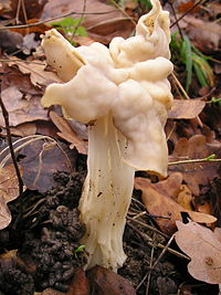
In the apothecium it can be seen that it is cup-shaped and when ripe it exposes the hymenophore where the asci are found. An example of this structure can be found in Pezizales as Helvella brevis or Otidea grandis, in Helotiales as Mollisia undulato-depressula or in Sordariales as Lasiosphaeria hispida.
On the other hand, in the unilocular ascostroma or pseudothecium we find that the asci are formed immersed in locules and their wall is only made of stroma. In Pleospora herbarum we can find this structure.
For its part, the gymnotecia is a type of ascocarp formed by asci surrounded by intertwined hyphae. There are pathogenic species for humans that can develop this form, such as the dimorphic fungus Ajellomyces dermatitidis (Blastomyces dermatitidis, asexual stage) that generates blastomycosis.
And finally the casmothecium, which is a totally closed structure where the asci are organized in a basal way so that they are all at the same height and are released by breaking the wall. Examples of this type of ascocarp can be seen in Podosphaera fusca.
All these structures, in addition to containing the asci, can also be associated with an element called hamatecia that can be distinguished between paraphysis (elongated hyphae that originate at the base of the apothecia and perithecia), periphysis (short unbranched hyphae found inside of perithecia) and pseudoparaphysis (hyphae that originate above the asci of an ascostroma). In some cases we can find naked asci, as in the mold Eremascus fertilis.
The type of asci, the arrangement they present, as well as their relationship with sterile structures of an ascocarp (hamatecia) are some criteria of taxonomic importance for the classification of this group of fungi.
Phylogeny
Ribosomal genetic analysis reveals the following relationships for subdivisions and classes of Ascomycetes:
| Ascomycota |
| |||||||||||||||||||||||||||||||||||||||||||||||||||||||||||||||||||||||||||||||||||||||||||||||||||||||||||||||||||||
Taxonomy
Ecology
Ascomycetes play a central role in most terrestrial ecosystems. They deal with the decomposition of organic materials, such as dead leaves, stems, fallen trees, etc. and they help detritus-eating animals that live on organic matter to obtain their nutrients. They process materials such as cellulose and lignin, which are difficult to exploit, for all this they play a very important role in the natural cycles of nitrogen and carbon.
The fruiting bodies or ascocarps provide food for a diverse set of animals, such as insects, slugs, snails, rodents and large mammals such as deer and wild pigs.
Ascomycete fungi are also known for their symbiotic relationships with other organisms.
Lichens
Probably from very early on, ascomycetes “domesticated” Chlorophyta algae or green algae, as well as other types of algae and cyanobacteria. Together they form mutualistic relationships known as lichens, which subsist in extremely inhospitable regions of the earth, including the arctic, deserts and high mountain tops, and can withstand extreme temperatures between -40 °C to +80 °C. While the photoautotrophic partner, the alga, creates metabolic energy through photosynthesis, the fungus offers support and protects against radiation and dehydration. About 42% of Ascomycetes (approximately 18,000 species) form lichens and the vast majority of lichen-forming fungi belong to this group. The proportion of basidiomycetes is two or four percent.
Mycorrhizae and endophytic fungi
Ascomycetes form two important types of relationships with plants: mycorrhizae and endophytes. Mycorrhizae are symbiotic associations of the fungus with the root system of plants; the fungus absorbs salts and minerals from the soil much more efficiently than the roots of the plant. For its part, the plant provides the products of photosynthesis to the fungus. In the case of many species, such as most conifers and many other plants, this association is of vital importance. In some cases, the fungus even transports nutrients from one plant to another, contributing to the robustness of the ecosystem. Mycorrhizae may have existed very early in the process of invasion of terrestrial environments by plants. There is evidence that the oldest land plant fossils already had mycorrhizae.
The endophytes live inside the plants, especially in the stems and leaves, but generally do not harm the host. The exact nature of this relationship is not yet fully understood, but it appears that this association confers increased resistance against insects, nematodes, and bacteria; it is also possible that it contributes to the production of toxic alkaloids used by plants in their defense against herbivores.
Symbiotic Relationships with Animals
A number of species of Ascomycetes of the genus Xylaria are found in the nests of South American leafcutter ants and other fungus-cultivating ants of the Attini tribe and also in the fungus cultures of the termites (Isoptera). These fungi only form ascocarps after the insects have left, so it is thought that these are fungi cultivated by them compared to what occurs in several cases of associations with Basidiomycota.
Bark beetles, in the family Scolytidae, are important symbionts of ascomycetes. The females transport the spores to the new host plant in sacs, called mycetangia, under the chitin. They burrow tunnels into the wood and make chambers or cells that they use to lay their eggs. When they lay their eggs they also leave spores from which hyphae grow that can effect the decomposition of the wood. When the larvae hatch they feed on the fungi. After metamorphosis they carry spores with which they can infect other trees. A well-known example of this is the Dutch elm disease, caused by the fungus Ophiostoma ulmi, transmitted by the elm bark beetle Scolytus multistriatus
Order Endomicetales
It's the yeast. saccharomycetaceans. They do not produce hyphae. They are in charge of some fermentations (see: Louis Pasteur), some species live anaerobically: Saccharomyces cerevisiae and Saccharomyces ellipsoideus; converting the sugars into ethyl alcohol and carbon dioxide. Reproduction is asexual by budding, during the formation of buds the nucleus undergoes division and one of the daughter nuclei passes to the new bud. Saccharomyces cerevisiae is heterothallic with + and - strains, then plasmogamy (fusion of the cytoplasm) occurs followed by karyogamy (fusion of nuclei), then the diploid (2n) cell becomes ascus and after undergo meiosis, four ascospores are formed, there is only diploid and haploid budding in Saccharomyces cerevisiae.
Class Euascomycetes
They produce sporocarps. The first phase occurs with plasmogamy but by not undergoing karyogamy, its genetic load remains "n+n" (binucleated). Mitosis occurs and then karyogamy occurs. The hyphae are septate (with divisions) regularly, there is presence of chitin, and there is also a central pore that interconnects the hyphae with each other.
Contenido relacionado
Molecular biology
Bromus
Amaryllidoideae


