Aorta
The aorta is the main artery of the human body, it measures an average of 2.5 cm in diameter in adults. It originates in the left ventricle of the heart, its initial trajectory is ascending, later it forms an arch called the aortic arch and descends through the thorax until it reaches the abdomen, where it divides into the 2 common iliacs that go to the lower limbs. It transports and distributes oxygen-rich blood and gives rise to all the arteries of the system circulatory except the pulmonary arteries that arise in the right ventricle of the heart.
Features
The aorta is an elastic artery that starts from the upper portion of the left ventricle, its wall is flexible and extensible. When the left ventricle of the heart contracts in systole and forces blood into the aorta, it expands. This stretch produces the potential energy that helps maintain blood pressure during diastole, during which time the aortic wall retracts.
Arterial compliance is the ability of blood vessels to distend and contract in response to changes in volume and pressure. The distension of the initial portion of the aorta propagates along the arterial walls in the form of a distension wave that reaches the small peripheral arteries and constitutes the pulse wave. It can be easily detected by palpation of the radial artery at the wrist.
Parts of the aorta
The aorta is divided into three parts: ascending aorta, aortic arch or aortic arch, and descending aorta. The latter is divided into two parts separated by the diaphragm called the thoracic aorta and the abdominal aorta.
Ascending aorta
It is the first portion of the aorta, about two inches long, part of the left ventricle of the heart with which it communicates through the aortic valve. As soon as it leaves, it leans forward and to the right, passing in front of the pulmonary artery, at its very origin it gives off the first two branches, the left and right coronary artery, which are of crucial importance, since they supply oxygen and nutrients to the heart. It presents a dilation at its beginning called the bulb of the aorta, which corresponds to the external visualization of the aortic sinuses (or of Valsalva ), from which the coronary arteries originate.
Aortic arch
The aortic arch, also called the aortic arch, lies between the ascending and descending aorta. Its central or proximal portion is shaped like an inverted u and gives rise to the brachiocephalic trunk, the left common carotid, and the left subclavian artery. At the midpoint of this arch or arch, the aorta passes from the anterior mediastinum to the posterior mediastinum. In the wall of the aortic arch, a large number of pressure-sensitive nerve terminals are located, which are called baroreceptors and are of great importance for the maintenance of blood pressure. Close by are the aortic bodies, which are groups of cells that form small structures about 2 mm in diameter that contain chemoreceptors capable of detecting changes in the composition of the blood, especially the concentration of oxygen.
Descending aorta
It is the section that goes from the aortic arch to the place where it divides into the common iliac arteries. On its way it crosses the diaphragm that differentiates it into two regions: thoracic aorta and abdominal aorta.
Thoracic Aorta
This is the name given to the half of the descending aorta that goes from the end of the aortic arch to the diaphragm.
Abdominal aorta
This name is given to the half of the descending aorta that spans from the diaphragm to its bifurcation.
Histology
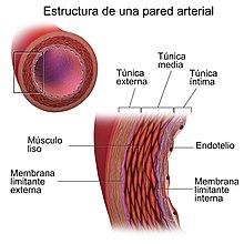
The wall of the aorta is formed by three concentric layers: intima, media, and adventitia. The intima is in direct contact with blood and is made up of endothelial cells. The media is much thicker, made up of up to 50 concentric layers of elastin sheets interspersed with contractile smooth muscle cells. The adventitia is the outermost layer, it is relatively thin and is made up of fibroblasts, collagen and elastic fibers.
The aorta and its branches
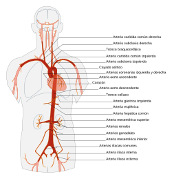
The ascending aorta gives off two branches, the coronary arteries, which distribute blood to the myocardium. It then turns to the left side of the body, where it forms the arch of the aorta (aortic arch), which descends and ends at the level of the intervertebral disc that separates the fourth and fifth thoracic vertebrae.
Continuing its descent, it becomes the descending aorta, approaches the vertebral bodies, crosses the diaphragm, traverses the abdomen, and divides at the level of the fourth lumbar vertebra into the primitive iliac arteries, which carry blood to the lower limbs. Throughout its route it emits vessels that branch into smaller ones that distribute the blood in the different organs. In these, the arteries divide into arterioles, and then into capillaries, which supply all the tissues of the body, except the pulmonary alveoli.
- Branches of the ascending portion: coronary arteries.
- Cayate branches of the aorta (Aortic Arc): brachyophalic trunk, common left carotid artery and left subclavian artery.
- Branches of the descending portion.
- Branches of the thoracic descending portion: bronchial arteries, esophageal arteries, mediastinal branches of the thoracic aorta and subsequent intercostal arteries.
- Branches of the abdominal descending portion:
- Parietal branches: lower diaphragmatic artery and lumbar arteries;
- Visceral branches: celiac trunk, upper mesenteric artery, medium adrenal artery, renal artery, gonal arteries (ovarian artery/testicular artery), and lower mesenteric artery.
- Terminal branches: middle sacra artery, primitive arteries right and left.
| Portion and branches |
Aorta ascendente
|
Cayado de la aorta
|
descending chest
|
Abdominal aorta
|
Pathologies
- Aorta aneurysm. It is a dilation located in a sector of the artery wall.
- Aortic coartation. It consists of a localized narrowing of the artery.
- Aortic dissection. It is a tear from the arterial wall that allows the blood to flow between its layers and force them to separate, forming a false light on the wall.
- Traumatic break. It can be by serious injury or trauma, for example, throbbing and crushing the chest. It's a very serious situation with high mortality.
- Transposition of the large vessels. It's a congenital disease in which the artery aorta comes out of the right ventricle instead of the left ventricle.
- Atherosclerosis. It consists of the deposit of lipidic substances in the inner layer of the arterial wall.
- Long-running aortic. It consists in the formation of an ulceration on a previous lesion by arteriosclerosis, penetrating the wall to a certain depth.
- Acute aortic syndrome. It is not a disease in itself, it can be due to penetrating ulcer, intramural hematoma or aortic dissection. Symptoms start acutely, highlighting chest pain.
Contenido relacionado
Fur
Medulla oblongata
Abdomen
Shoulder
Anencephaly


