Anatomy
Anatomy is a science, a branch of biology, that studies the structure of living beings, that is, the shape, topography, location, arrangement and relationship among themselves of the organs that it's made of. It can be classified into descriptive, functional and surgical anatomy. Anatomy is based above all on the descriptive examination of living organisms, however the understanding of this architecture implies also studying the function, which is why it is related to physiology and is part of a group of basic sciences called morphological sciences (biology). of development, histology and physical anthropology), which complete his area of knowledge. Human anatomy is one of the basic or preclinical sciences of medicine. The scientist who cultivates this science is called an anatomist, the Dictionary of the Spanish language of the Royal Spanish Academy also accepts the term anatomical.
Etymology
Derived from Latin. anatomĭa, and from the Greek. ἀνατομία [anatomy]; derived from the verb ἀνατέμνειν [anatémnein], 'to cut' or 'separate' made up of ἀνά [aná], 'upward' and τέμνειν [témnein], 'to cut')
Subdivisions
- Descriptive or systematic anatomy. Study the different systems independently. Systematic anatomy can be divided into several sciences:
- Neuroanatomy. Study the nervous system.
- Myology. Study muscles.
- Osteology. Study the bones.
- Arthrology. Study the joints.
- Spolacnology. Study the different viscera, especially those contained in chest, abdomen and pelvis.
- Topographic anatomy. Also called surgical anatomy or regional anatomy. It is the branch of the anatomy that studies the relationships between organs and structures located in a particular region. It therefore describes organisms by regions in which there may be parts that belong to different systems. In topographic anatomy when describing an area is usually done from the most superficial to the deepest, so in a region typical of the lower or higher human member there would be a foreground formed by the skin, then the subcutaneous cell tissue followed by a superficial aponeurosis. More in depth we will find one or two muscle planes and in the deeper limit a bone structure formed by a bone. It should be borne in mind that this description is of a general nature and there are numerous variations depending on the specific area to be considered.
- Clinical anatomy. Study those aspects of the structure and function that are useful in the practice of medicine.
- Comparative anatomy. Study the similarities and differences between the structures of organisms belonging to different species.
- Microscopic anatomy or histology. It is the branch of the anatomy that studies the tissues using the microscope.
- Macroscopic anatomy: It is dedicated to the study of organs or parts of the body large enough to be able to observe them in plain sight, without using microscope.
- Anatomy of development. Also called embryology, it is responsible for studying the development of organisms from the moment of conception to birth.
- Functional anatomy: Study the morphology of the different organs and their relationship with the function they perform.
- Surface anatomy. It is the study of the anatomical structures that can be seen from the outside of the organism. It includes the determination of a series of reference points on the surface that correspond to the projection of structures in depth.
- Radiological anatomy: Study the form through images obtained by simple radiology, computed axial tomography (CT) or ultrasound.
- Pathological anatomy. It is the branch of medicine that studies the morphological bases of the disease.
- Artistic anatomy: Study anatomical art.
- Plant anatomy: or phytotomy is the field of the Botanic that studies the internal structure of the plants.
- Animal anatomy: Also called veterinary anatomy, describes the morphological structures of the different animals.
- Human anatomy: Describes the morphological structures of the human body.
Related Sciences
Anatomy is related to other related sciences, including the following:
- Embryology: Study the embryonic development of different organisms.
- Histology: Study the structure of organs and tissues from a microscopic point of view.
- Physiology: Study the function of organs and systems.
- Pathology. Branch of medicine responsible for the study of diseases.
- Paleoanthropology. It is the branch of physical anthropology that deals with the study of human evolution and its fossil record.
Nomenclature
Anatomical terminology has always been the subject of debate, given the large number of terms and the existence of synonyms and eponyms that can sometimes create confusion. For this reason, anatomists from different countries have agreed to establish a common, structured, systematic and universal terminology that is regularly updated in periodic meetings. It receives the name of International Anatomical Terminology and has been written by the International Federative Committee of Anatomical Terminology, it has been translated into different languages, including Spanish. It contains around 7500 terms that correspond to human macroscopic anatomical structures.
Anatomical plans
Anatomical planes are imaginary planes that cross the organism and divide it into two parts.
- Sagital plan. Cross the body longitudinally and divide it into a right and left half.
- Front plan. Also called a coronal plane, it crosses the body from top to bottom and divides it into a previous or frontal half and a posterior or dorsal
- Transverse plan. It is a plane that crosses the body from the front backwards and divides it into a upper and lower half.
Addresses
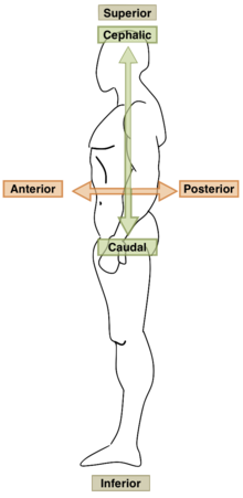
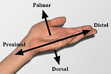
For the study of anatomy, anatomical directions can be used to indicate relative locations. The address base of the human being is very similar to that of the rest of vertebrates. Vertebrates are integrated into this study methodology thanks to the basic arrangement they share: The main anatomical directions are:
- Cephalic or superior. Located at the top of the body.
- Caudal or lower. When placed at the bottom of the body
- Medial. Located on the middle line or near the middle line.
- Lateral. Located away from the middle line.
- Ventral or earlier.
- Dorsal or later.
- Proximal. Located near its origin.
- Distal. Located away from its origin.
- Surface. Located on the outside of the body.
- Deep. Located in the inner part of the body.
- Aferente. It is said of what goes from outside to inside, or from the periphery to the center.
- Efferent. It is said of what goes from inside out, or from the center to the periphery.
- Rostral. It refers to the part of an organ or structure that is closer to the face.
- Palmar. It refers to the palm of the hand.
- Plant. It refers to the sole of the foot.
In tetrapod vertebrate animals such as the horse, the standard anatomical posture is one in which the animal stands upright and faces forward. In this form it is dorsal superior, ventral inferior, cephalic in front and caudal in back.
Fish Anatomy
The anatomy of fish is determined by the physical characteristics of water, much denser than air, with less dissolved oxygen and greater light absorption, and by the evolutionary component of each species within the superclass Pisces.
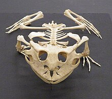
Amphibian Anatomy
Amphibians are gill-breathing tetrapods in the larval stage. In the adult phase they lose their gills and develop lungs, the circulation becomes double, they present a heart with three chambers formed by a ventricle and two auricles.
Reptile Anatomy
Reptiles present a set of morphological adaptations common to most of the representatives of the class. Among them the presence of an exoskeleton provided with epidermal horny scales, paired limbs with five fingers (except in snakes), well-ossified skeleton, presence of lungs which allows them to live on dry land, a heart with three chambers, two auricles and a ventricle (except in crocodiles that have four chambers), internal fertilization and shell-covered eggs.
Bird Anatomy
The anatomy of birds presents many adaptations designed to make flight possible. Birds have evolved to possess a light and powerful skeletal and muscular system which, together with the circulatory and respiratory systems, makes them capable of very high oxygenation and metabolic activity. Due to these anatomical specializations, they have been assigned a class of their own in the Chordate Edge.
Mammalian Anatomy
In mammals the body is divided into three sectors: head, trunk and tail, although this may be missing, for example in the gorilla, or transformed into a fin as in the whales, in some species the tail is prehensile and can be used as limb. There are four extremities on the trunk that may be adapted to walking, digging, grasping objects, flight (bats) or swimming (cetaceans). The females present mammary glands to feed the newborns with milk. The circulation is double and complete, the heart consists of four chambers, as in birds, two atria and two ventricles. Breathing is pulmonary. The digestive system presents in some groups such as ruminants special adaptations. Fertilization is internal, embryonic development usually takes place inside the maternal uterus thanks to the placenta, although in some groups the uterus is rudimentary and development is completed in a marsupial bag, as occurs in metatheria. In some species such as monotremes (platypus) embryonic development takes place in an egg, outside the maternal organism.
Human Anatomy
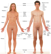
Human anatomy has many similarities to that of most mammals. For your better understanding it is divided into systems and devices.
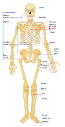
Skeletal System
Also called the skeletal system, it is formed in a human adult by a total of 206 bones. It represents about 12% of the weight of an adult. Bones are attached to each other at joints and are closely linked to ligaments, tendons, and muscles. The human skeleton is divided into two parts:
- Axial skeleton, formed by the skull, spine, ribs and stern. It consists of 80 bones.
- Appendicular skeleton, formed by the bones of the upper and lower limbs along with the escapular and pelvic waist. It consists of 126 bones.
- Escape tape. Clax and omoplate.
- Senior member. Húmero, cúbito, radio, carpos, metacarpos,falanges
- Girl tape. Pelvis
- Lower member. Fémur, throtula, tibia, perone, tarso, metatarso, falanges.
Muscular system
It is made up of voluntary muscles. Its function is to achieve the movement of the body. The set of all skeletal muscles corresponds to 40% of the weight of an average adult.
Nervous system
The nervous system consists of the brain, spinal cord, and peripheral nerves. Nerves can be sensory that carry information from the periphery to the brain, motor that carry signals from the brain to the muscles, and mixed that have both sensory and motor fibers. The main cell is the neuron that is endowed with extensions that join it to other neighboring neurons, forming a complex network of interconnections. The nervous system collects information from the outside world through the senses or internally from the different organs, processes it, and emits signals through the motor nerves that cause muscle contraction and movement. It is also involved in the production of hormones by the different glands (neuroendocrine system).
Circulatory system
The circulatory system or cardiovascular system is made up of a fluid called blood, a set of tubes (arteries, veins, capillaries) and a driving pump that is the heart. It is an internal transport system that serves to move nutritive elements, metabolites, oxygen, carbon dioxide, hormones and other substances within the body. The human circulatory system, like that of all mammals, is double. The right side of the heart sends deoxygenated blood to the lungs through the pulmonary artery, the already oxygenated blood is directed through the pulmonary veins to the left side of the heart, which distributes it throughout the body through the aorta artery and its branches. Blood is produced in the red bone marrow, it is made up of blood plasma and the formed elements that are platelets, red blood cells (erythrocytes) and white blood cells (leukocytes).
Digestive system
The digestive system is made up of the digestive tract and a set of accessory structures. The digestive tract begins in the mouth and continues through the pharynx, esophagus, stomach, small intestine, large intestine, and anus. The accessory structures are a set of glands that discharge their secretions into the digestive tract, including the salivary glands that produce saliva, the pancreas, the liver, and the biliary system that transports bile to the duodenum.
Food passes from the mouth down the esophagus to the stomach. In the stomach, the food is mixed with an acid secretion and proteolytic enzymes to form a semi-liquid paste called chyme. The stomach contents are emptied into the duodenum, where bile produced by the liver and pancreatic juice from the pancreas are added. Nutrients are absorbed in the small intestine, then the large intestine absorbs mostly water and salts. minerals. Eventually, the remaining substances pass into the rectum and are excreted through the anus.
Respiratory system
The respiratory system is the set of organs used to exchange gases between the environment and the blood. It is made up of the larynx, trachea, bronchi and lung.
Urinary system
It is a set of organs responsible for the production, storage and expulsion of urine. It is formed by two kidneys, two ureters, the urinary bladder, and the urethra.
Male reproductive system
It is made up of the testicles, epididymis, prostate, vas deferens, urethra, and penis.
Female reproductive system
Composed of the ovaries, fallopian tube, uterus, vagina, and vulva.
Endocrine System
The endocrine system is the set of organs and tissues in the body that secrete a type of substance called hormones. Hormones are chemical messengers released by cells that reach the bloodstream to remotely regulate different bodily functions, including growth rate, tissue activity, metabolism, and the development and function of the sexual organs. Once the destination point is reached, these mediators bind to their specific receptor located on the target cell. The major hormone-producing glands are the thyroid gland, which produces thyroxine, the pancreas, which produces insulin, the pituitary gland, which secretes numerous hormones, and The adrenal glands that produce cortisol, aldosterone, and adrenaline.
Exocrine glands
The exocrine glands are a group of glands that are distributed throughout the body and produce non-hormonal substances. They are part of different systems, some of the most important are the parotid gland that produces saliva, the mammary gland that produces milk, the lacrimal gland that produces tears, and the sweat glands that produce sweat.
Sensory System
The sensory system is the part of the nervous system responsible for processing information from the sense organs. It is made up of sensory receptors and those parts of the brain involved in signal processing. The main sensory systems are: vision from receptors located in the eye, hearing from receptors located in the inner ear, taste from receptors located in the tongue, smell from receptors located in the nasal cavities, balance from receptors located in the inner ear, and touch. from receptors located in the skin.
Integumentary System
It is made up of the skin, nails, hair, sweat glands and sebaceous glands. The skin constitutes a barrier that defends the body from external physical and chemical agents and prevents the invasion by bacteria and viruses. It is divided into several layers, the outermost is called the epidermis and the deeper dermis.
Main structures
| Equipment and systems | Main structures | |
|---|---|---|
| Digestive device | Esophagus, stomach, small intestine, large intestine, liver, pancreas, gallbladder. | |
| Circulatory fitting | Artery heart (pulmonary artery, aorta artery), veins (pulmonary veins, superior vena cava, lower vena cava), blood capillaries. | |
| Respiratory device | Faringe, larynx, trachea, lung. | |
| Nervous system | Brain (brain, cerebellum, spinal cord, nerves). | |
| Endocrine system | Hypophysis, thyroid, parathyroids, adrenal glands, pancreas, ovary, testicle. | |
| Muscle system | Sternocleidomastoid, deltoids, trapeze, major pectoral, wide dorsal, brachyal biceps muscle, brachial triceps muscle, abdominal rectum, external oblique muscle of the abdomen, major gluteal muscle, larger aductive muscle, quadiceps muscle, sural triceps, ischiotibile muscles. | |
| Bone system | Skull, lower jaw, spine, ribs, stern, scapula, collarbone, humerus, cubit, radio, hand bones, pelvis, femur, tibia, perone, foot bones. | |
| Urinary appliance | Kidney, urinary bladder, ureters. | |
| Male genital appartment | Testicular, deferential duct, prostate, urethra, penis | |
| Female genital apparel | Ovary, fallopian tube, uterus, vagina, vulva. | |
| Immune system | Bone medulla, spleen, lymph nodes, thymus | |
| Tegumentary system | Skin, hair, nails, sweat glands |
History of Anatomy
- Old age
Anatomy as a science took its first steps in Ancient Greece. Herophilus of Chalcedon (335 BC-280 BC), physician of the Alexandrian school, was one of the first to practice dissections of human corpses. Erasistratus (304 BC – 250 BC) distinguished the main structures of the brain and observed that the nerves converged towards the central nervous system.
Galen of Pergamum (Pergamum, 129-Rome, c. 201/216), was a Greek physician and philosopher who lived in the Roman Empire. He produced an immense corpus, distributed in more than 125 volumes, dealing with the anatomical-functional study of various systems: muscular, nervous, respiratory, and circulatory. His preponderance as a teacher of medicine lasted for more than 1,400 years. This knowledge also continued to be taught during the Middle Ages. However, Galen's statements contained numerous errors that were accepted without criticism, many of them derived from extrapolating structures observed in animals to the human species.
- Age
The anatomical knowledge of the Middle Ages is based on the acceptance of Galenic anatomy. The classes taught by the professor were made with the lectio of Galen's text, the few dissections in cadavers were carried out by a practitioner while reading the classic, without criticism.
- Modern Age
Galenic Medicine begins to be questioned from the anatomy. Andrés Vesalio, considered the father of modern anatomy, dedicated himself to the dissection of cadavers to obtain anatomical knowledge. He recorded his observations in his De humani corporis fabrica.
- Contemporary Age
The appearance of the microscope opened up a new microscopic descriptive world, microscopic anatomy or histology, and the gradual conversion of anatomy into dynamics from Vesalius's static fabric, incorporating function and relationship into his observations.
Artistic Anatomy
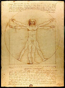
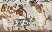
The representation of the human body does not depend solely on knowledge of anatomy, since the different artistic styles often do not seek realism. The paintings of Ancient Egypt, for example, show a series of conventions that adopt the real anatomy at the convenience of the artist. On the contrary, in Ancient Greece morphology was meticulously studied and the aim was to represent the human figure in its maximum perfection. During the Renaissance, many famous artists studied anatomy from life and even practiced dissections themselves to improve the representation of the body including Michelangelo, Raphael, Dürer, and Leonardo da Vinci.
Anatomical Associations
- International Federation of Anatomist Associations
- Pan American Anatomy Association
- Central American and Caribbean Anatomy Association
- International Journal of Morphology
- Pan American Academy of Anatomy
Contenido relacionado
Kingdom (biology)
Homo sapiens
Asexual
