
Adipose tissue
The adipose tissue or fatty tissue is tissue of mesenchymal origin (a type of connective tissue) made up of the association of cells that accumulate lipids in their cytoplasm: the adipocytes.
Adipose tissue, on the one hand, fulfills mechanical functions: serving as a shock absorber, protecting and keeping both internal organs and other external structures of the body in place, and metabolic functions: generating fats for the body.
Types of adipose tissue or fatty tissue
There are two types of adipose tissue, white (or unilocular) adipose tissue and brown (or multilocular) fat.
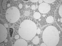
The cytosol and nucleus are reduced to a small area near the membrane. The rest is occupied by a large blob of fat. Adipose tissue, which lacks ground substance, is divided by fine trabeculae of fascicular tissue into lobules.
The fat in the cells is in a semi-liquid state and is also composed mainly of triglycerides. It accumulates preferentially in the subcutaneous tissue, the deepest layer of the skin. Its cells, lipocytes, are specialized in forming and storing fat. This layer is called the adipose panicle and is an insulator from cold and heat. It acts as a pad and also as a storehouse of nutritional reserves.
This type of tissue fills and cushions, especially in the subcutaneous areas. It also serves as structural support and an energy reserve function. Fat varies in consistency, that is, it can be found in both a liquid and solid state.
The growth of this tissue can be produced by cell proliferation (hyperplastic growth), by accumulation of a greater amount of lipids in existing cells (hypertrophic growth), but the number of adipocytes never increases by mitotic division. During adolescence, growth is generally rapid, the same as in the hypertrophic adult.
Anatomical features
Humans

In humans, adipose tissue is located under the skin (subcutaneous fat), around internal organs (visceral fat), in the bone marrow (yellow bone marrow), and in the breasts. Adipose tissue is located in specific regions, which are known as adipocyte stores. In addition to adipocytes, which make up the highest percentage of cells in adipose tissue, there are other cells that are collectively present called the visceral stromal fraction (SVF). This stroma is formed by preparatory-adipocytes, fibroblasts, adipose tissue macrophages, and endothelial cells. Adipose tissue contains small blood vessels.
In the integumentary system, which includes the skin, fat tissue is stored in the deepest layer of the skin regulating body temperature.
Around organs, this tissue provides protection. However, its main function is to be a reserve of lipids, which can be used to generate the necessary energy for the body and protect us from excess glucose. Under normal conditions, it provides hunger and satiety stimulation to the brain.
Mice

Mice have eight large deposits of adipose tissue, four of which are located in the abdominal cavity. Paired gonadal reservoirs are attached to the uterus and ovaries in females and to the epididymis and testes in males. Of all the deposits in mice, the gonadal deposits are the largest and the easiest to dissect, comprising approximately 30% fat.
Paired deposits of the retroperitoneum are found along the dorsal wall of the abdomen, surrounding the kidney and sometimes extending to the pelvis.
The deposit of the mesentery forms a network of tissue that supports the intestines; and lastly, the omental reservoir, which originates near the stomach and spleen, when abnormally enlarged it extends to the abdomen. Both reservoirs store both lymphoid tissue and lymph nodes and white blood cells respectively.
The two superficial deposits are the inguinal paired deposits, which lie anterior to the upper extremities (under the skin); includes the group of lymph nodes in the groin.
The subscapular deposits are not a mixture of brown adipose tissue and white adipose tissue, found under the skin between the dorsal ridges and the scapulae. The layer of brown adipose tissue in this reservoir is normally covered by a "glaze" of white adipose tissue; sometimes these two types of fat (brown and white) are hard to tell apart. Minor reservoirs include the pericardium, which surrounds the heart, and the paired popliteal reservoirs, located in the popliteus muscle (behind the knee), each popliteal reservoir containing a large lymph node.
Obesity
In people who are morbidly obese, the excess fatty tissue that hangs down from the abdomen is referred to as the panicle. Complicating morbidly obese surgery, the panicle remains literally a “skin apron” if a severely obese person loses large amounts of fat too quickly (a common result of gastric bypass surgery). This condition cannot be effectively corrected by diet and exercise alone, since the panicle is made up of adipocytes and other types of supporting cells shrunk to their minimal volume and diameter. Reconstructive surgery is one of the most viable ways to treat it.
Obesity and cancer
According to the International Agency for Research on Cancer, and based on epidemiological studies, people who are obese or overweight have a higher risk of developing several types of cancer, such as adenocarcinoma of the esophagus, colon cancer, breast cancer (in postmenopausal women), endometrial cancer, and kidney cancer.
Stomach fat

Visceral or abdominal fat also known as intra-abdominal fat, is located within the abdominal cavity, stored between organs (stomach, liver, intestines, kidneys, etc.). Visceral fat is different from subcutaneous fat, located under the skin, or intramuscular fat, which is scattered throughout skeletal muscles. Fat in the lower body, such as the thighs and buttocks, is subcutaneous and not a space-aware tissue, while fat in the abdomen is more visceral and in a semi-liquid state. Visceral fat is composed of by adipose deposits, including mesenteric tissue and white adipose tissue of the epididymis, and perirenal deposits. Visceral fat is considered as adipose tissue while subcutaneous fat is not considered as such.
Excess visceral fat is known as central obesity, which protrudes from the abdomen. It has also been linked to type 2 diabetes, insulin resistance, inflammatory diseases, and other obesity-related diseases.
The female sex hormone causes fat to be stored in women's thighs, buttocks, and hips. Men are more likely to have fat stored in the belly due to the hormonal difference that exists. When women reach menopause and ovarian estrogen production declines, fat migrates from the thighs, buttocks, and hips to their waists, where it is then stored in their belly.
High intensity exercise is an effective way in which abdominal fat can be reduced. Another study shows that at least 10 hours a week of energy expenditure through aerobic exercise is required for fat reduction abdominal.
Epicardial fat
Epicardial adipose tissue (EAT) is a particular form of visceral fat deposited around the heart and recognized as an active organ of metabolism that generates several bioactive molecules, which can significantly affect cardiac function. Marked component differences have been observed between epicardial fat and subcutaneous fat, thus suggesting the existence of a storage depot of fatty acids that specifically impact adipocyte functions and metabolism.
Subcutaneous fat

Much of the remaining non-visceral fat is found just under the skin in a region called the hypodermis. This subcutaneous fat is unrelated to some of the classic obesity-related pathologies, such as heart disease, cancer, and cerebrovascular accident (CVA); and some evidence even suggests that it may have a protective function. The typical pattern of female body fat distribution (or pelvis) around the hips, thighs, and legs is subcutaneous fat, and it is for this reason that it represents a lower health risk. compared to visceral fat.
Like other fat organs, subcutaneous fat is an active part of the endocrine system, as it secretes the hormones leptin and resistin.
The relationship between the subcutaneous adipose tissue layer and total body fat in a person is normally modeled using regression equations. The most popular of these equations was formulated by Durnin and Wormersley, who rigorously tested many types of skin folding and, as a result, created two formulas for calculating the body density of women and men. These equations present an inverse correlation between skin folds and body density—as the sum of skin folds increases, body density decreases.
Factors such as sex, age, population size, economy, among other variables can invalidate and make these equations unusable; Until 2012, the Durnin and Wormersley equation remains only to estimate the true level of fat in a person. New formulas are still being created.
Physiology
Free fatty acids are released from lipoprotein by an enzyme called lipoprotein lipase; these free fatty acids enter the adipocyte, where they are reassembled into triglycerides. Approximately 87% of human fat tissue is made up of lipids.
There is a constant flow of free fatty acids. These fluids are controlled by insulin and leptin. If we have a high concentration of insulin there is an increase in the flow of free fatty acids, when insulin drops, fatty acids can be released from adipose tissue. The secretion of insulin is stimulated by the elevated concentration of sugar or glucose in the blood due to the consumption of carbohydrates.
In humans, lipolysis (hydrolysis of triglycerides into fatty acids) is regulated by the controlled balance of lipolytic androgen-B receptors and α2A-androgen receptors, mediating anti-lipolysis. Adipocytes have an important physiological role in regulating triglyceride and free fatty acid levels, as well as determining insulin resistance.
Abdominal fat has a different metabolism, being more likely to induce insulin resistance. This explains why central obesity is a precursor of glucose intolerance, being an independent factor in cardiovascular diseases (even in the absence of diabetes mellitus and hypertension).
In 2009 studies were conducted on female monkeys at Wake Forest University, which found that individuals who suffered from high levels of stress had higher levels of visceral fat in their bodies. This suggests a possible cause-effect where stress promotes the accumulation of visceral fat, which becomes the cause of hormonal and metabolic changes that contribute to cardiovascular disease and other health problems. Recent advances in biotechnology have allowed the harvesting of adipose tissue from adult cells, allowing stimulation of the tissue using the patient's own cells. In addition to this, it is known that cells differentiated into adipocytes, both from humans and animals, can be reprogrammed to be pluripotent cells without the need for culture cells. The use of the patient's own cells reduces the risk of rejection and avoids ethical problems. associated with the use of stem cells from an embryo.
Adipose tissue is a great source of aromatase, which in both men and women contributes to the production of estradiol. Hormones derived from adipocytes include:
- Adiponectin
- Resistant
- Plasminogen activator and inhibitor (PAI-1)
- TNF T
- IL-6
- Leptina
- Estradiol
Adipose tissue also secretes a type of cytokines, called adipokines, which act on the complications associated with obesity.
Brown Fat
Brown adipose tissue is a specialized form of adipose tissue in humans, rodents, small mammals, and some hibernating animals. It is found mainly around the neck and in large blood vessels in the chest. This specialized tissue can generate heat by "uncoupling" of the respiratory chain of oxidative phosphorylation within the mitochondria. The uncoupling process refers to when protons down the electrochemical gradient pass through the inner mitochondrial membrane, the energy from this process is released as heat instead of being used to generate ATP. This thermogenic process may be vital in newborns exposed to cold, who in turn require this thermogenesis to maintain heat, since they are unable to shiver, or perform other actions to stay warm.
Attempts to simulate this process pharmacologically have so far been unsuccessful (and even lethal). Techniques to manipulate the differentiation of “brown fat” have become a mechanism for weight loss therapy. in the future, promoting the growth of tissue with this specialized metabolism without inducing it in other organs.
Until recently, brown adipose tissue was thought to be limited primarily to the infant stage in humans, but new evidence has overturned that belief. Metabolically active tissue exhibits similar temperature responses to brown adipose tissue, this was first recorded in the neck and trunk of some human adults in 2007, and the presence of brown adipose tissue in human adults was subsequently verified. histologically in the same anatomical areas.
Genes
The thrifty gene hypothesis (also known as the starvation hypothesis) states that in some populations, the body would be more efficient at retaining fat in times of plenty, thereby endowing it with greater resistance to starvation in times of abundance. of food shortages. This hypothesis has been discredited by physical anthropologists, physiologists, and the original proponent.
In 1995, Jeffrey Friedman, in his residence at Rockefeller University, discovered leptin, a protein that the genetically obese mouse lacked. Leptin is produced in white adipose tissue and by signaling to the hypothalamus. When leptin levels drop, the body interprets it as a loss of energy, and hunger increases. Mice lacking this protein eat up to four times their normal size.
Leptin, however, plays a different role in dietary obesity in rodents and humans. Because adipocytes produce leptin. Leptin levels are elevated in obesity. However, hunger remains, and, when leptin levels drop due to weight loss, hunger increases. The fall in leptin is better viewed as a signal of hunger than the increase in leptin as a signal of satiety. However, the elevation of leptin in obesity is known as leptin resistance. The changes that occur in the hypothalamus to give rise to leptin resistance in obesity are currently the focus of obesity research.
Genetic defects in the leptin (ob) gene are rare in human obesity. As of July 2010, only 14 people from five families have been identified worldwide as carrying a mutated ob gene (one of which was the first identified cause of genetic obesity in humans): two families of Pakistani origin living in the UK, one family living in Turkey, one in Egypt and one in Austria. Two other families have been found to carry a mutated obreceptor. Others have been identified genetically as partially deficient in leptin, and, in these individuals, leptin levels at the lower end of the normal range can predict obesity.
Several gene mutations involving melanocortins (used in brain signaling associated with appetite) and their receptors have also been identified as causing obesity in a larger portion of the population than leptin mutations. In 2007, researchers isolated the fat gene, the researchers hypothesized that this gene serves to keep animals lean during times of plenty. In that study, increased adipose gene activity was associated with leaner animals. Although its discoverers named this gene the adipose gene, it is not a gene responsible for the creation of adipose tissue.
Physical properties
Adipose tissue has a density of about 0.9 g/mL (0.9 kg/L). Thus, a person with more fat tissue will float more easily than a person with the same weight, but with more muscle tissue, since muscle tissue has a density of 1.06 g/ml (1.06 kg/L).
Body fat meter
A body fat meter is a widely available tool used to measure the percentage of fat in the human body. Different meters use various methods to determine body fat, relative to weight. These tend to underestimate the percentage of body fat.
In contrast to clinical tools, a relatively inexpensive type of body fat meter uses the principle of bioelectrical impedance analysis (BIA) to determine an individual's percentage of body fat. To accomplish this, it passes a small, harmless electrical current through the body and measures the resistance, then uses the information about the person's weight, height, age, and gender to calculate an approximate value for percentage of fat. body of the person The calculation measures the total volume of water in the body (lean and muscle tissue contain a higher percentage of water than fat), and the percentage of fat is calculated based on this information. The result can fluctuate several percentage points depending on what has been eaten and the amount of water consumed prior to analysis.
Fonts
Genneser Finn. Histology. (2666). Panamerican Medical Editorial. Third edition. Buenos Aires, Argentina, Colombia 1999 and Mexico D.F Chile, la serena (2011)
Contenido relacionado
Natural Sciences
Posidoniaceae
Calathea